A comprehensive exam is a vital portion of your eye health and is recommended at a minimum of every 2 years.
What components are part of a comprehensive eye exam?
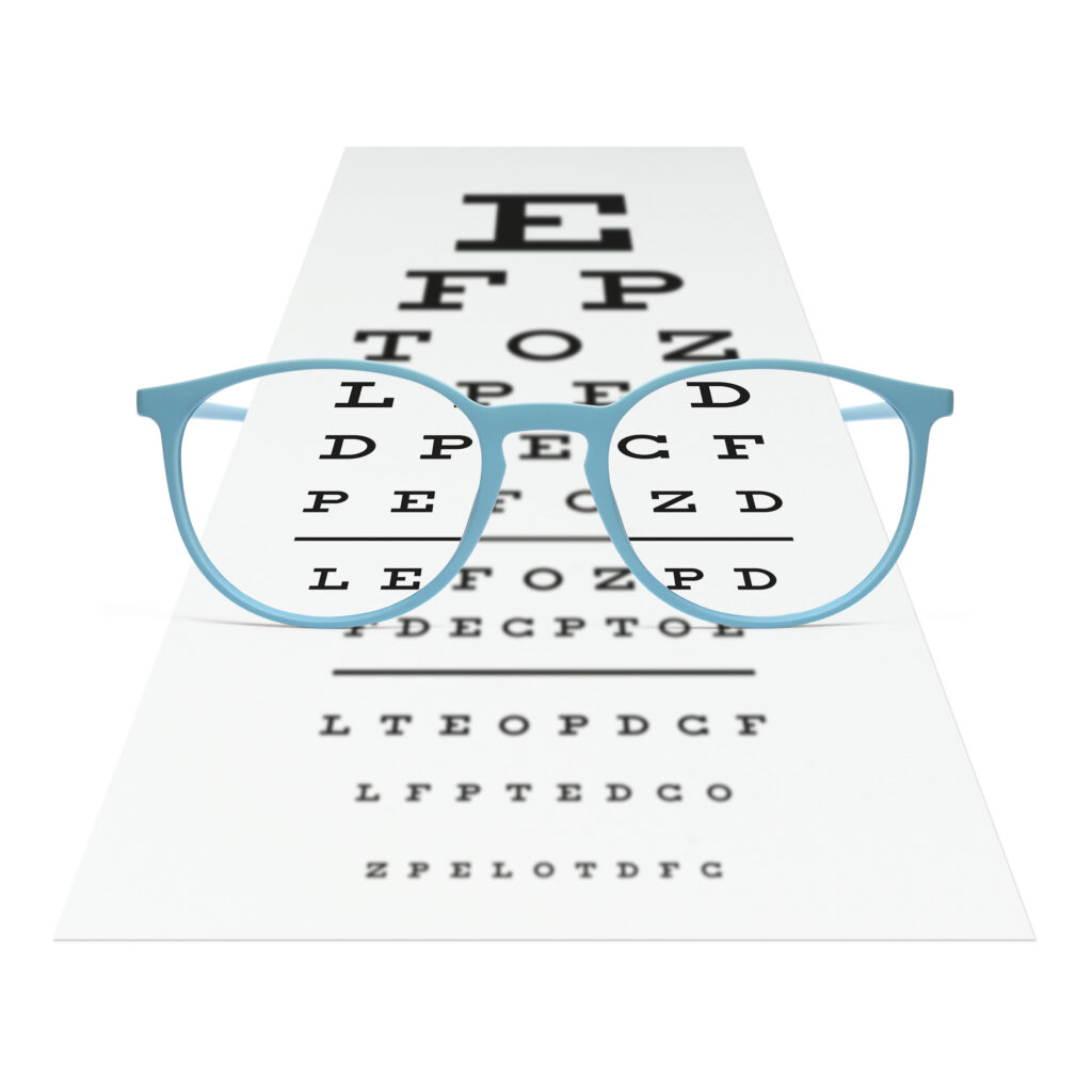
Visual acuity check
Visual acuity is the number value to your vision. You may have heard the term, “20/20 vision”. This refers to the ideal visual acuity of your eyes. Some individuals, however, have better vision at 20/15 and 20/10! If the visual acuity is worse than 20/20, it is often an indication of an underlying problem in the eyes.
Extraocular movements
We check your eyes’ ability to move in all directions. If there are any restrictions, it could point to neurological issues.
Eye alignment
We check the ways your eyes align at different distances with a test called the cover test. It lets us know whether you’re prone to more eye strain depending on where your eyes align.
Pupils
Checking pupils is a vital component to the eye exam that can point to subtle problems in the visual system. The test is performed by shining a light into your eyes
Peripheral/side vision
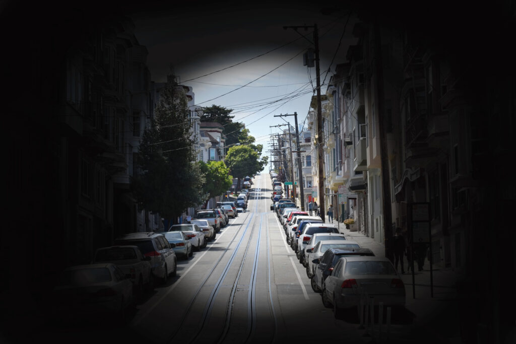
This image shows the field of view of a patient with Retinitis Pigmentosa. This is also seen in end-stage glaucoma.
We check your ability to see things outside of your central point of view. Often we can pick up on signs of stroke, genetic eye conditions and blood vessel problems in the eyes with a simple screening of your side vision.
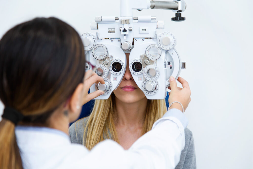
Refraction
This is the portion where we determine what prescription goes into your glasses, i.e. the classic “One or two” portion of the exam.
Ocular health
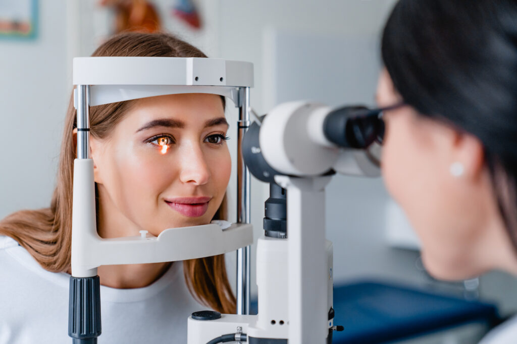
Slit lamp exam/Anterior segment exam
This portion is where we look at the front of your eyes and check for diseases of the front part of the eyes including signs of meibomian gland dysfunction, dry eye, corneal issues, cataracts…to name a few.
Intraocular pressure (IOP)
Testing the intraocular pressures or, in other words, the pressure inside the eye is one of the screening tests we perform for glaucoma. We utilize a machine called the iCare that easily gives us a reading of the eye pressure. We also utilize a test called Goldmann tonometry when necessary.
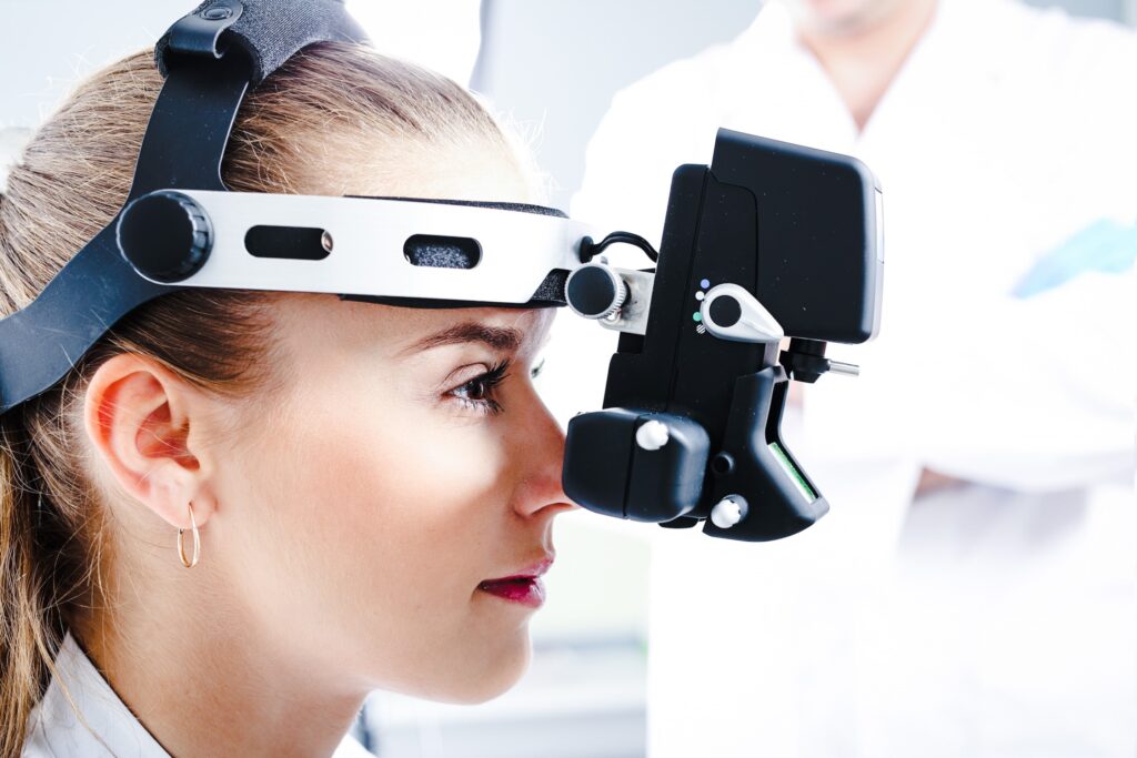
BIO/Posterior segment exam
We utilize a headlamp called binocular ophthalmoscope (BIO) and a magnifying lens to view the back of the eye. The back of the eye is best viewed when dilated. With this test we check for bleeds at the back of the eye related to systemic conditions such as diabetes and high blood pressure. We also check for tears, holes and thinning the retinal layer. In addition, we look for glaucoma and macular degeneration
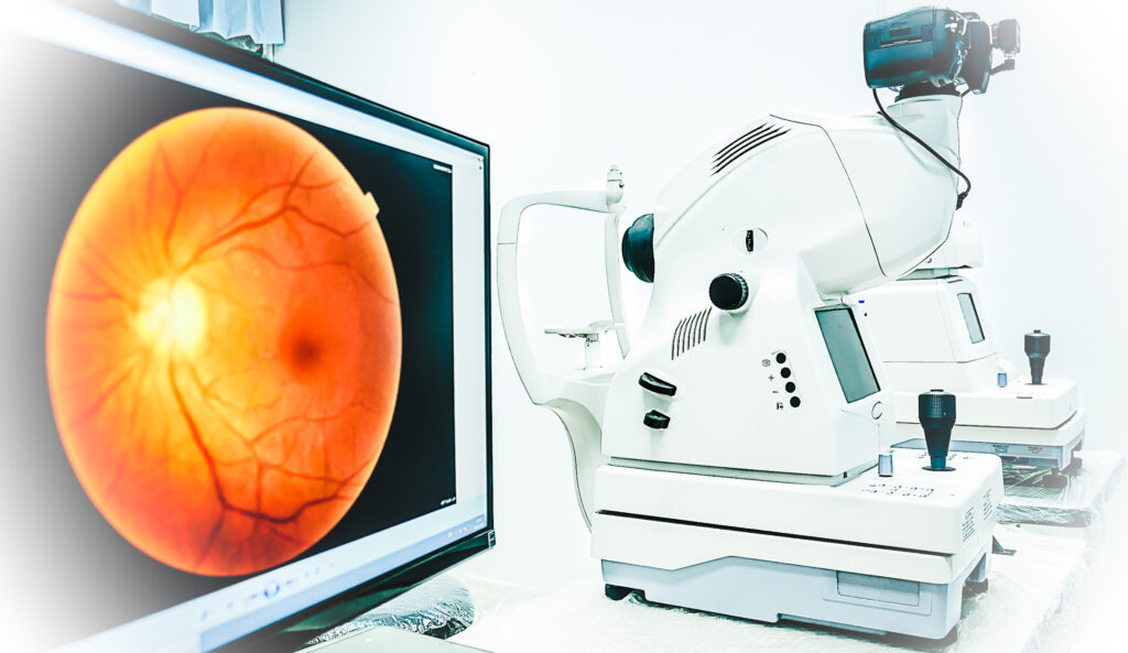
Dilation and photos of the back of the eye
As mentioned above, dilating the eyes is the best way to view the back of the eye and check for eye diseases. Dilation is recommended every 2 years.
In addition to dilating the eyes, taking a photo of the back of the eyes is strongly recommended. This allows to compare for changes over time! If all is healthy, photos are recommended every 2 years concurrently with dilation.